
Diagnostic imaging
We have recently fully-upgraded our imaging capabilities, with new, state-of-the-art x-ray and ultrasound systems.
This makes imaging a pleasure for us, and gives us high-resolution, crystal clear images, resulting in confidence in accurate and rapid diagnosis.

X-ray
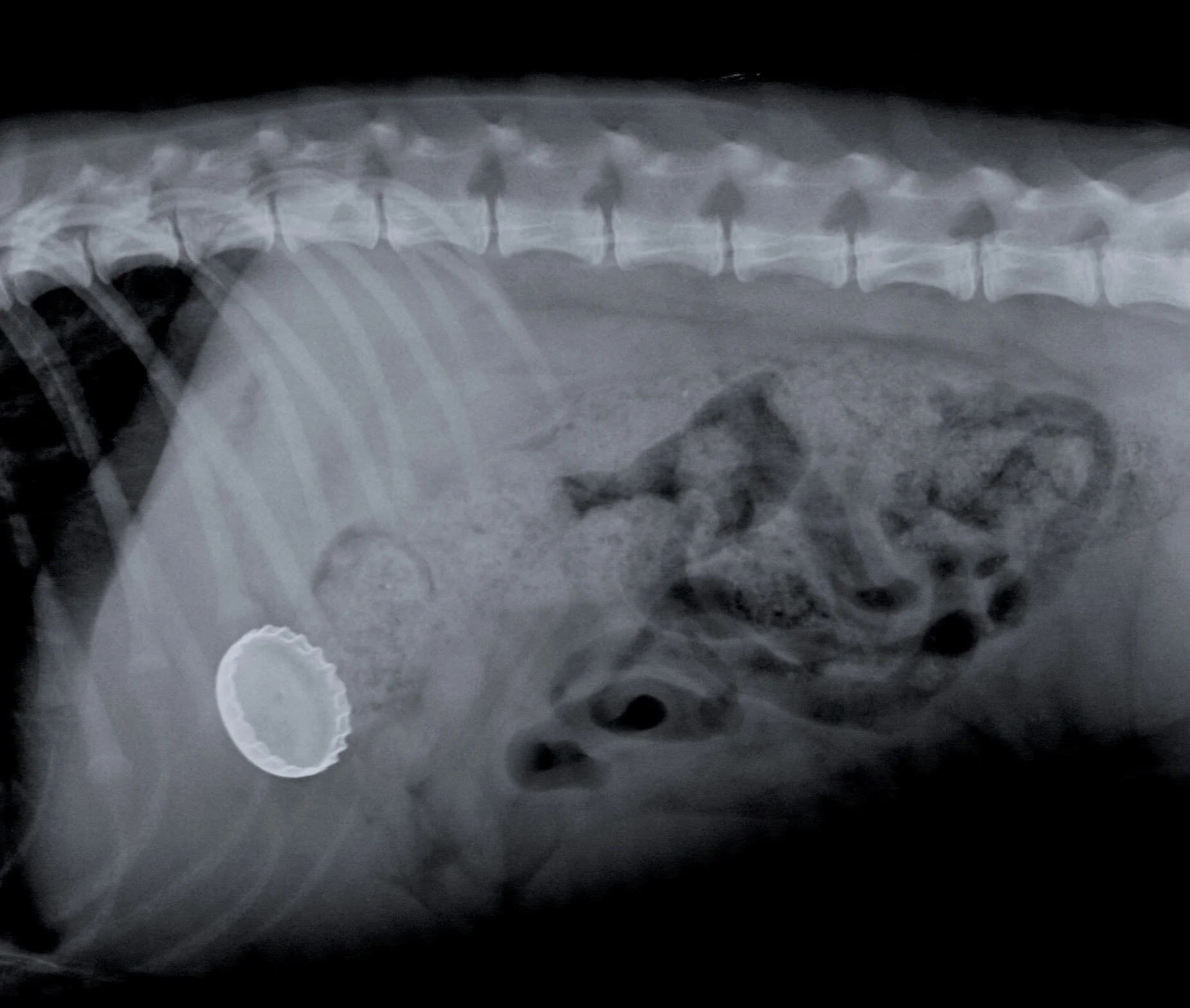
X-ray is the cornerstone of imaging in any veterinary practice, and we couldn’t live without it. X-rays can give us an exceptionally wide range of information.
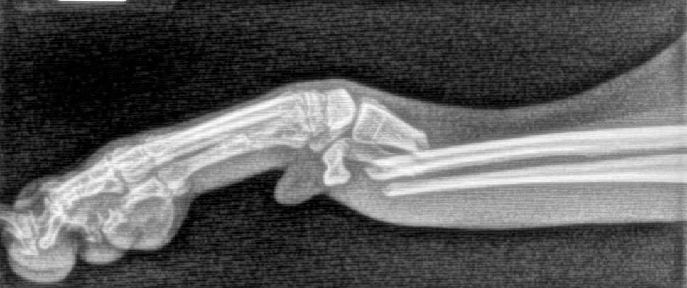
Broken bones
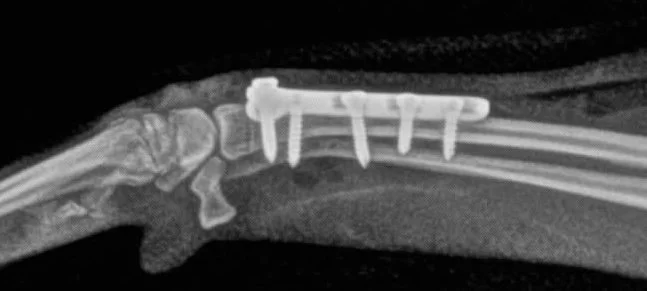
After repair
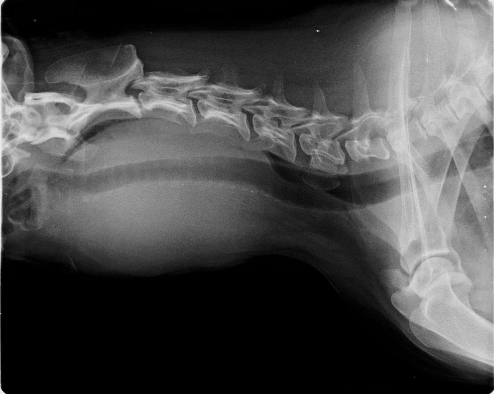
Cancer
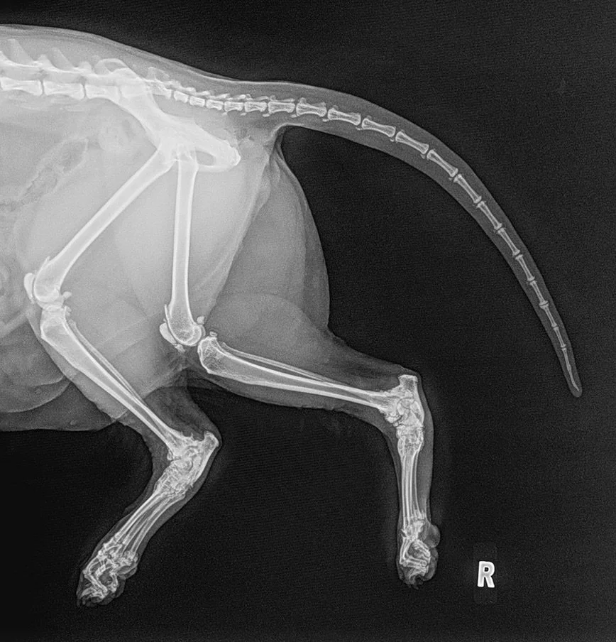
Arthritis
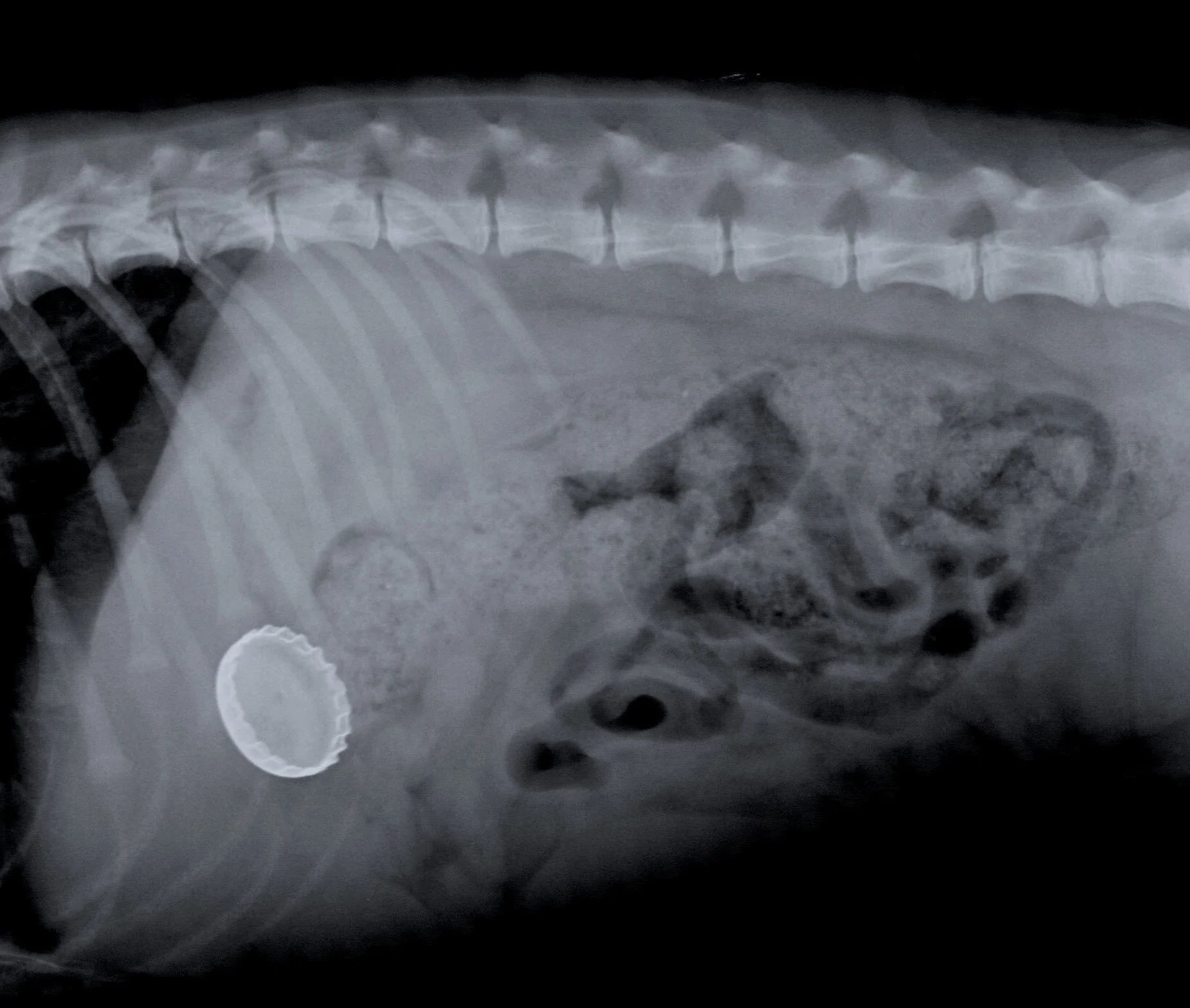
Foreign body
Our digital radiology platform allows rapid acquisition and exporting of a full diagnostic series of x-rays in just minutes, minimising time to reach a diagnosis, and time under sedation or anesthesia, when this is necessary.
It also allows us to give images to our clients, or forward them to radiology specialists anywhere in the world to assist us with interpretation in complex or unusual cases.

Ultrasound
Ultrasound is the other main imaging modality we use at Victoria. Its applications are different, but complementary to x-rays, and it’s quite common for us to use both modalities during a standard imaging work-up.
Ultrasound has the advantage of producing live images, in real time, without any radiation, so it’s extremely safe.
The main general drawback of ultrasound is that it’s technically challenging to perform, and takes years to master. Fortunately, all of the VVC vets have extensive experience with ultrasound, as well as advanced training and postgraduate qualifications.
Several of the key areas where we use ultrasound are:
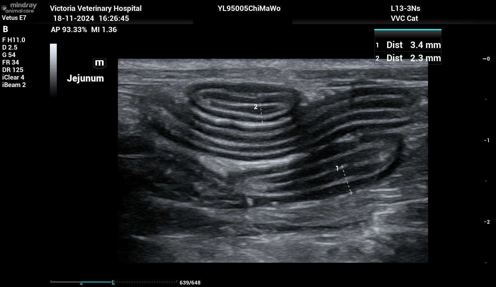
Abdominal ultrasound, where it is used to check all abdominal organs, including the liver, kidneys, bladder, intestines, spleen and pancreas, as well as checking for abnormalities like fluid buildup or cancers.
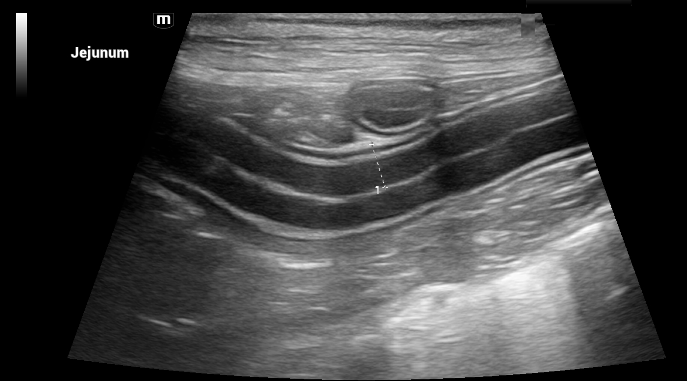
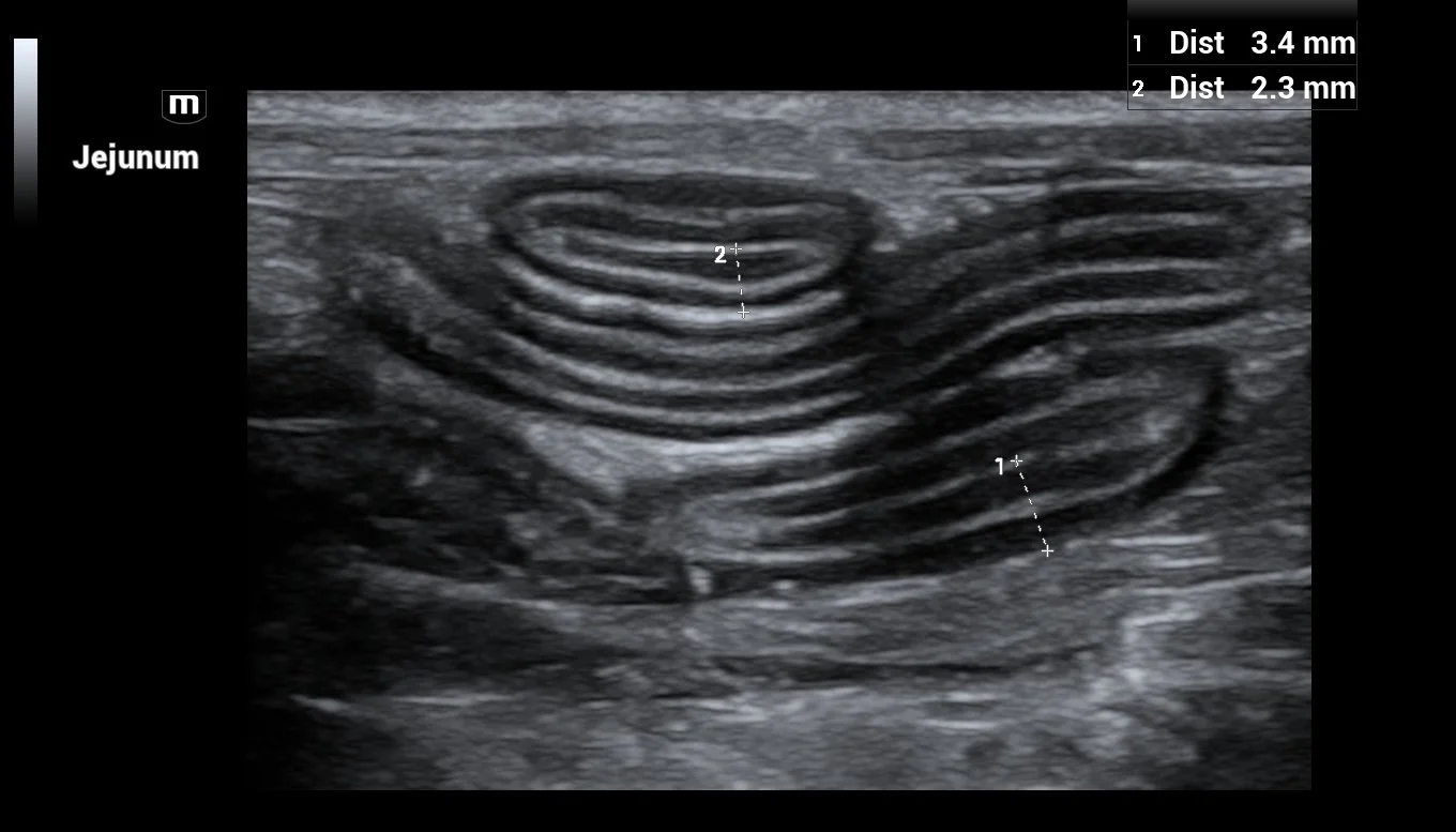
Normal vs abnormal (thickened) intestines, a key to diagnosis of intestinal disease.
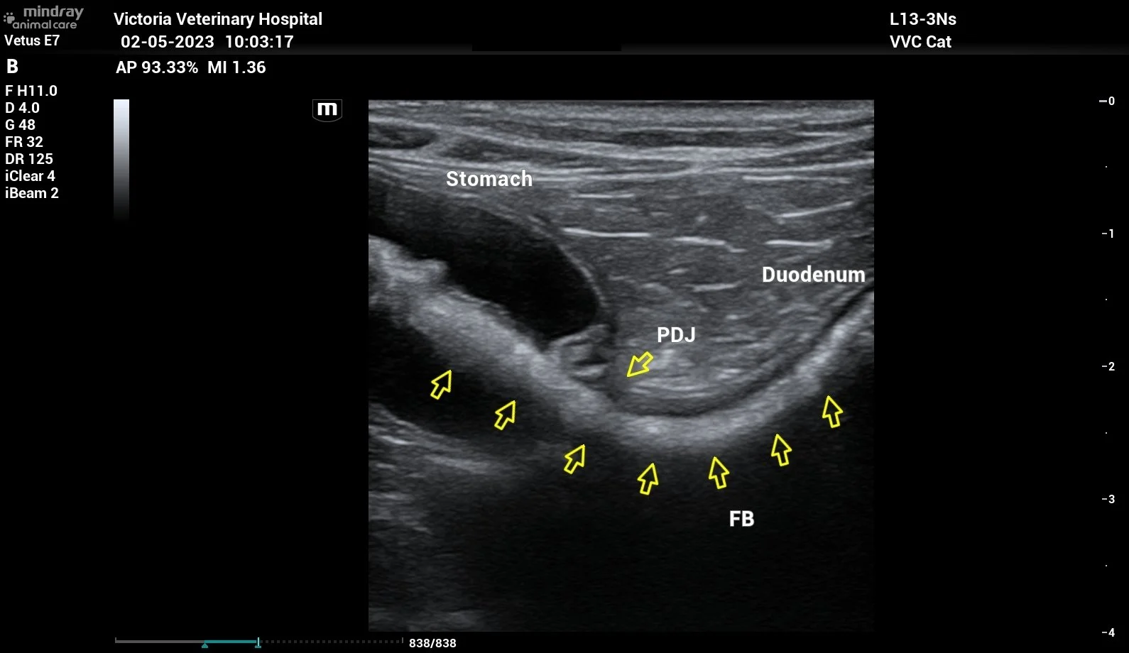
Ultrasound showing a linear foreign body (string) swallowed by a cat.
Cardiac ultrasound (also called echocardiography), which is used to examine the heart. We can use moving images to identify abnormalities within the heart.
Color-flow Doppler (CFD) ultrasound diagnosis of a leaking valve (mitral valve) in the heart.
…and we can use special tools to make measurements of flows and pressures within the heart, allowing us to determine the correct treatment, and monitor the effects of our treatments:
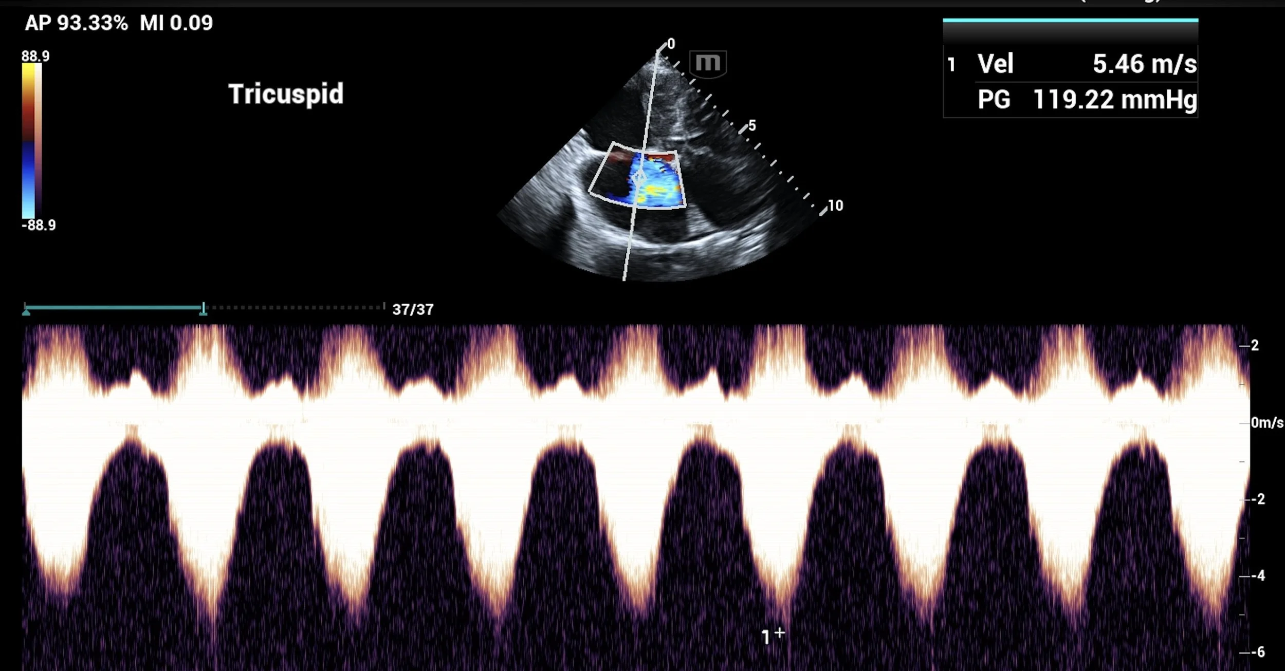
Spectral Doppler ultrasound diagnosis of pulmonary hypertension in a dog.
Ultrasound-guided diagnostic sampling: we can use ultrasound to guide biopsies of abnormal internal structures such as tumours, to allow quick and accurate diagnosis.
Ultrasound-guided biopsy of a mass in the abdomen.
Pregnancy diagnosis: sometimes we have the very enjoyable task of using the ultrasound to detect new puppies or kittens, and check that they are healthy!
A 45-day old puppy on a pregnancy scan.
Endoscopy
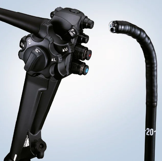
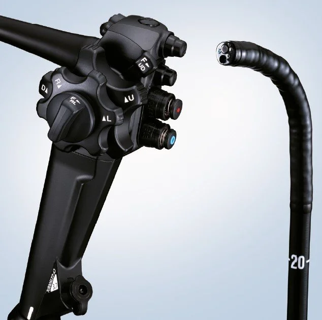
An endoscope is a camera connected via a fiberoptic cable to a flexible scope which can be used to look inside the body.
At Victoria we have a range of scopes suitable for various different purposes and anatomic sites.
Our largest scope is used for gastrointestinal endoscopy.We can use this for upper (esophagus, stomach and small intestine) and lower (colon) endoscopy.
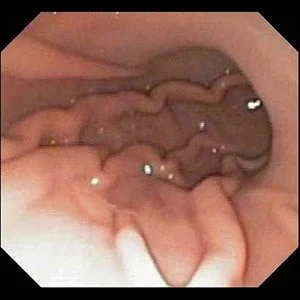
The scope can be used to check for abnormalities within these locations, and to take biopsy samples of abnormal tissues.
Endoscopy of the stomach
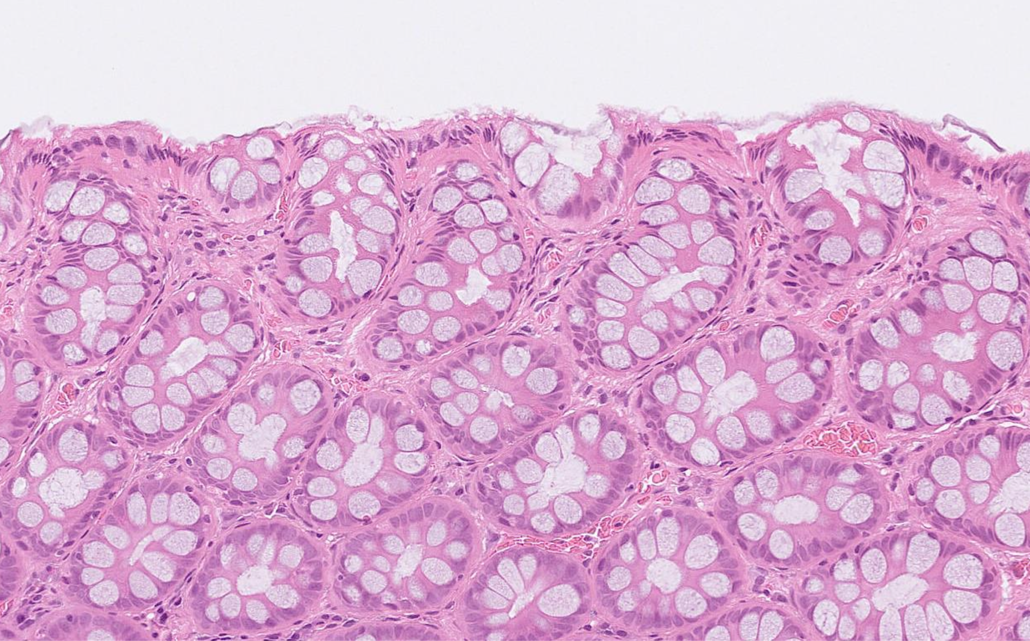
A biopsy of the colon viewed under the microscope.
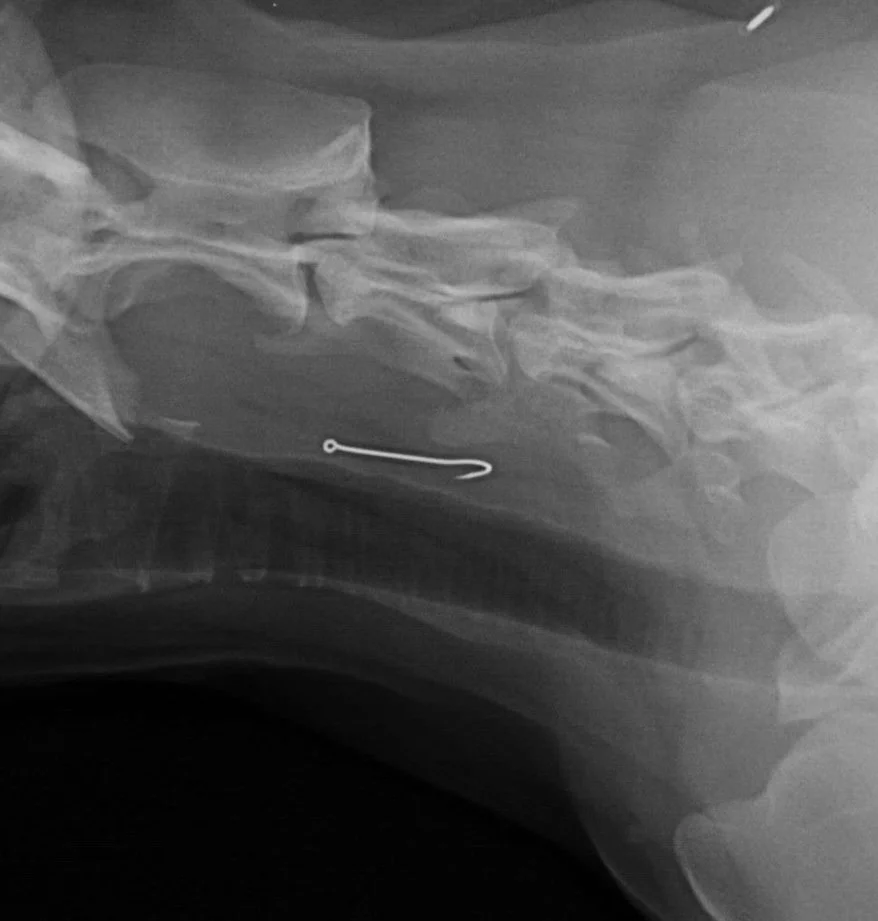
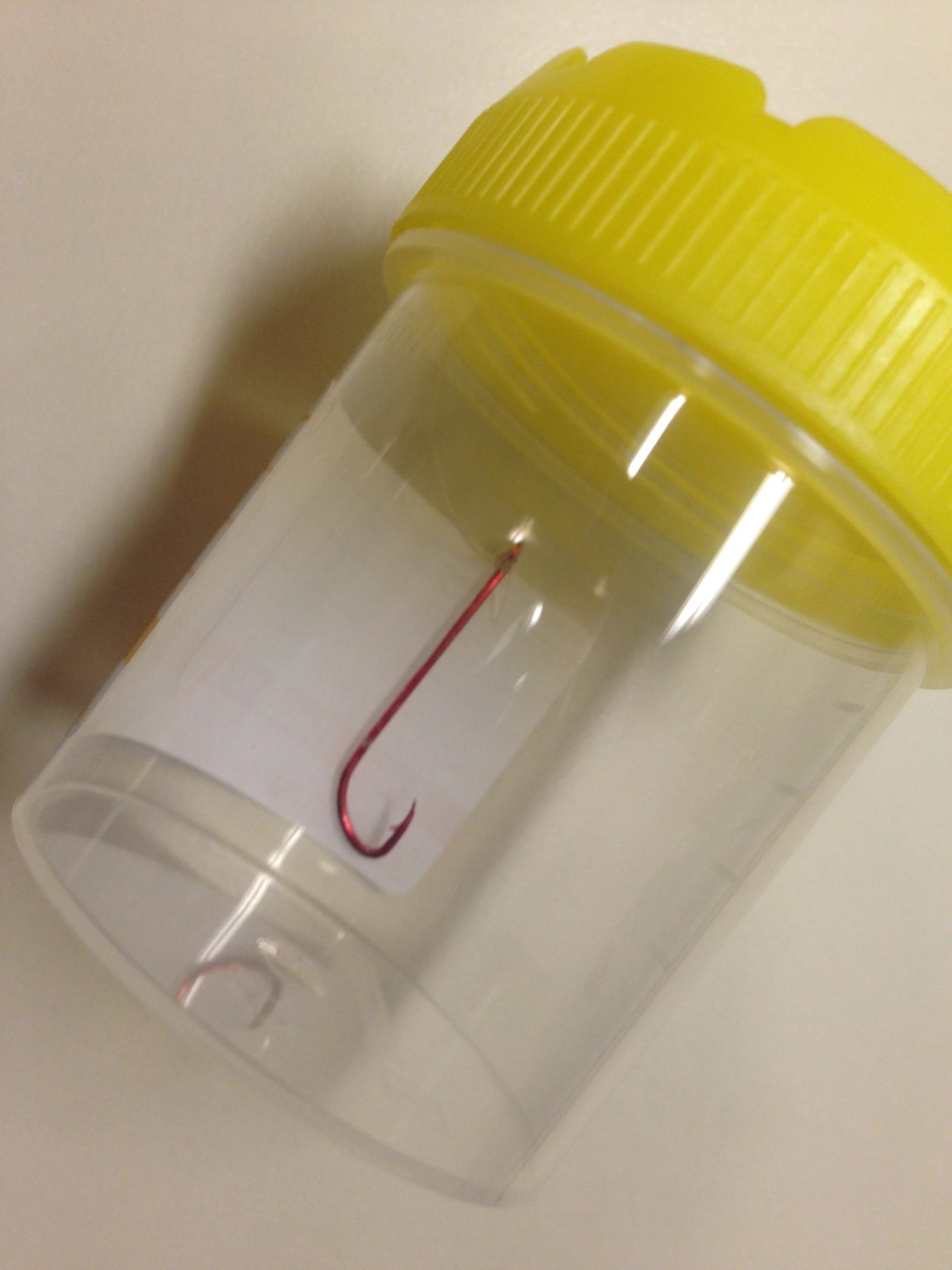
Fish-hook, swallowed by a dog, before and after endoscopic removal.
We also have a narrower scope small enough to fit into the airways. This is called bronchoscopy, and allows us to see inside individual airways while patients are under anesthesia, and take fluid or samples from abnormal sites.
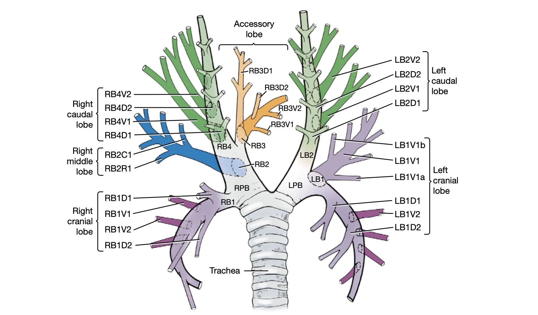
Map of the airways of a dog, which we can explore with bronchoscopy.
Bronchoscopy video of main airways under anesthesia.
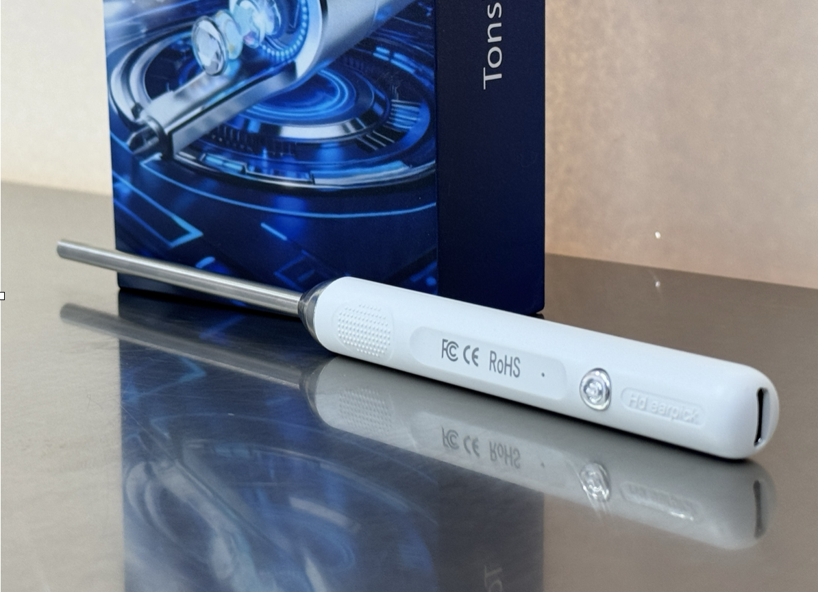
We also have a range of “pocket” scopes which can be paired with phones or tablets, and can be used for smaller sites such as ears and nose, including in the consult room to share ear examinations with clients in real-time.