

Logo for Victoria Veterinary Clinics featuring a stylized blue 'V' within a semi-circular shape, with the text "Victoria Veterinary Clinics" in English and Chinese, and "since 1992" underneath.

影像測驗

我們近期全面升級了影像設備,引進了全新的X光機和超聲波系統。

這不僅讓我們的影像檢測更加順暢高效,更提供了高解析度、更清晰的影像,使我們能更有信心地做出準確、快速的診斷。

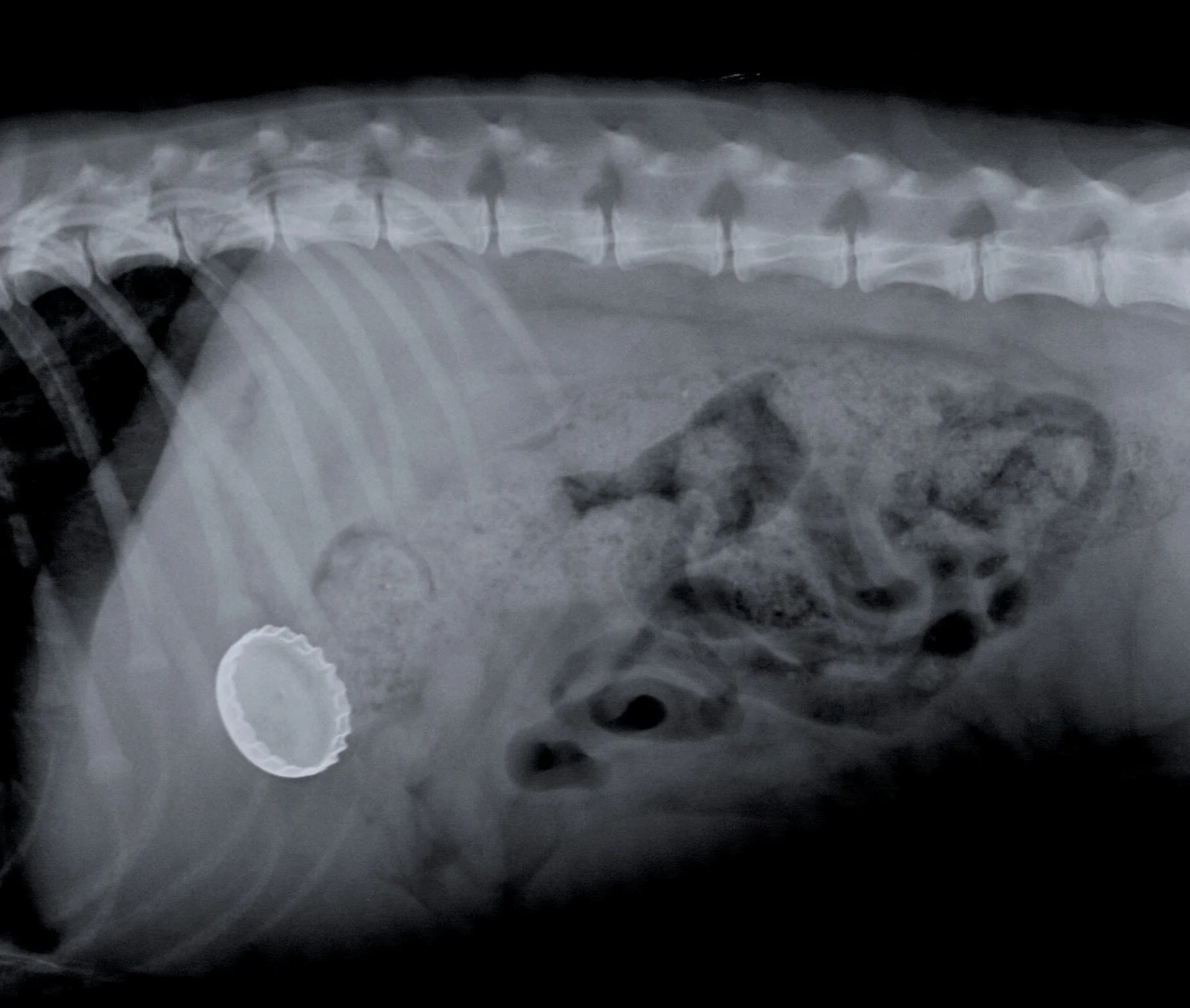
X-ray of a patient with a foreign body in the abdomen.
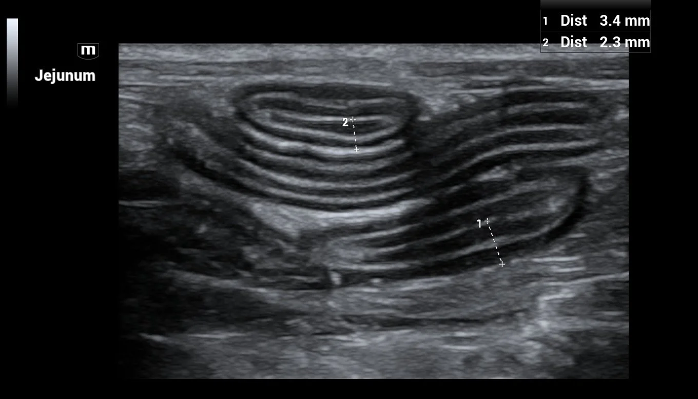
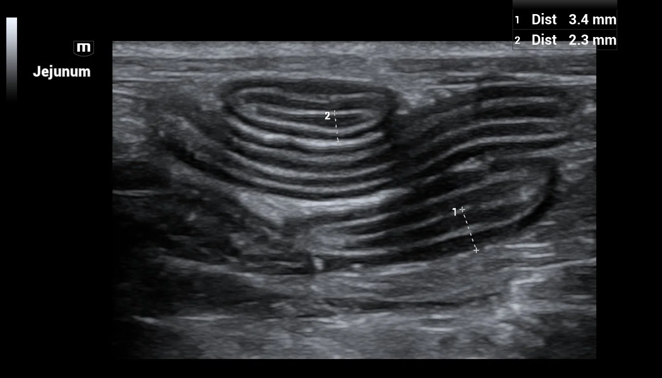
Ultrasound image showing multiple circular cross-sections of the small intestine, labeled as Jejunum, with measurements of 3.4 mm and 2.3 mm.
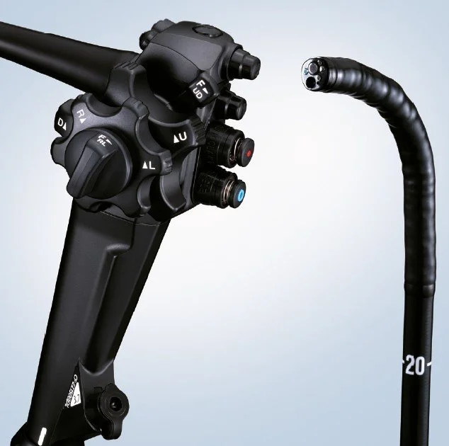
Close-up of a medical endoscope control handle with buttons and knobs, alongside a flexible black endoscope tube.

X光

X光檢查是任何獸醫診所影像診斷的基石,我們離不開它。 X光能為我們提供極為豐富的訊息,無論是…
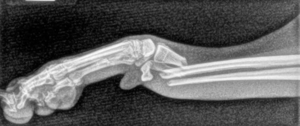
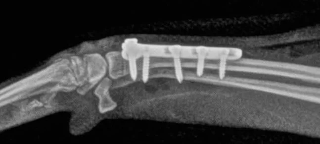
X-ray image of a dog front leg with visible broken bones.

骨折

X-ray image of a dog's forearm with surgical screws and a metal plate fixing broken bones.

修補後

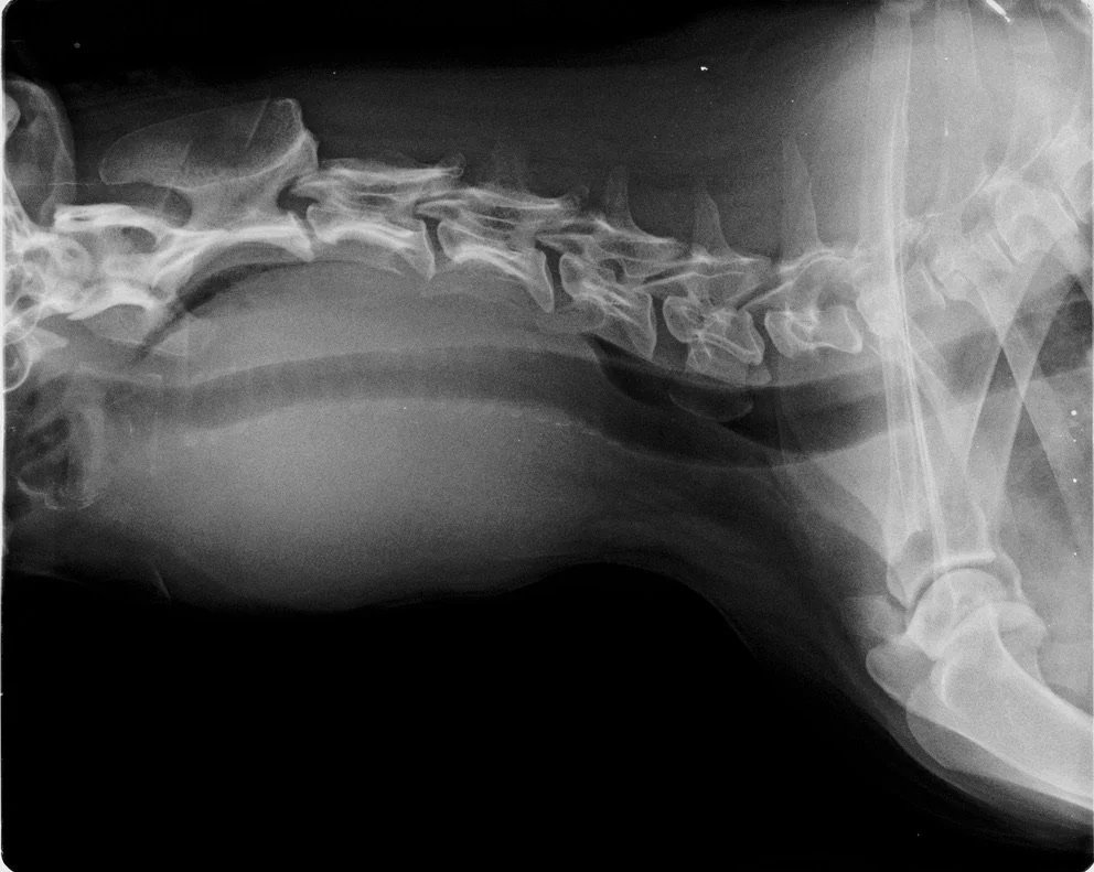
X-ray image of a dog's neck and upper chest, showing the tumour around the neck.

腫瘤

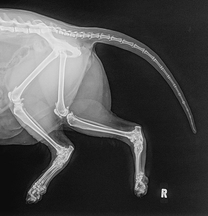
X-ray image of a small animal, likely a cat or dog, showing the spine, ribs, legs, and tail, with hind legs arthritis.

關節炎

我們的數位x光能夠快速取得並匯出完整的診斷級X光影像,從而縮短確診所需的時間;若需進行鎮靜或麻醉,也能減少動物處於此狀態的時長。

此外,我們還可以將影像資料提供給主人,或將其發送給世界各地的放射科專家,以便在處理複雜或罕見病例時獲得專業的閱片協助。
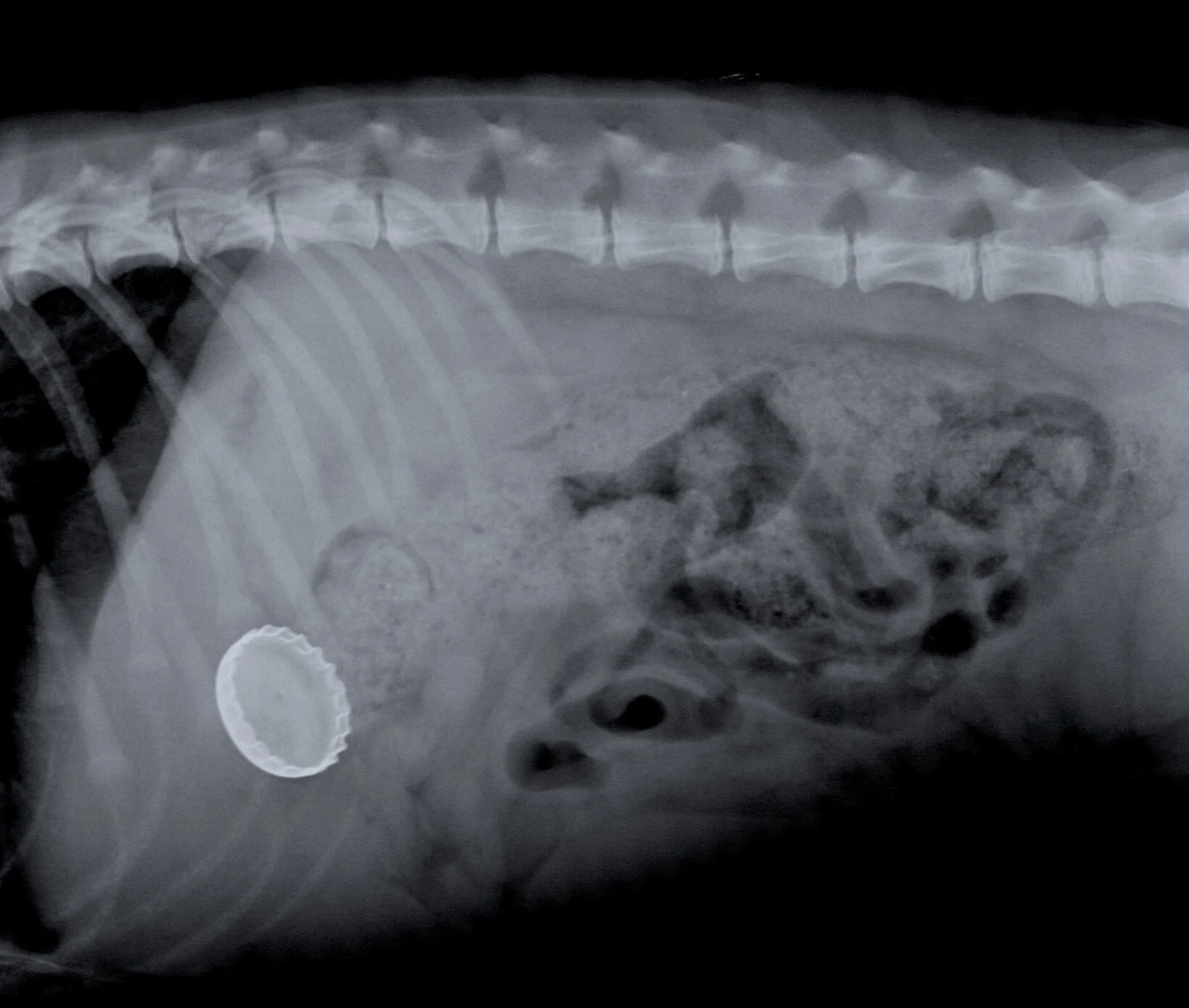
X-ray of a patient with a foreign body in the abdomen.

異物

超聲波

超聲波檢查是我們採用的另一種主要影像檢查。雖然其應用方式與 X 光不同,但兩者相輔相成;在常規影像檢查過程中,我們經常結合這兩種技術。

超聲波檢查的優點在於能夠即時產生動態影像,且不產生任何輻射,因此安全性極高。

超聲波檢查的主要限制在於其操作技術要求較高,需要多年的實踐才能熟練。幸運的是,維多利的獸醫不僅擁有豐富的超聲波檢查經驗,還接受過專業進階培訓並具備相關研究生資格。

我們應用超聲波檢查的主要領域包括:

腹部超音波檢查:用於檢查肝臟、腎臟、膀胱、腸道、脾臟和胰臟等所有腹部器官,並檢查積水或腫瘤等異常病變。
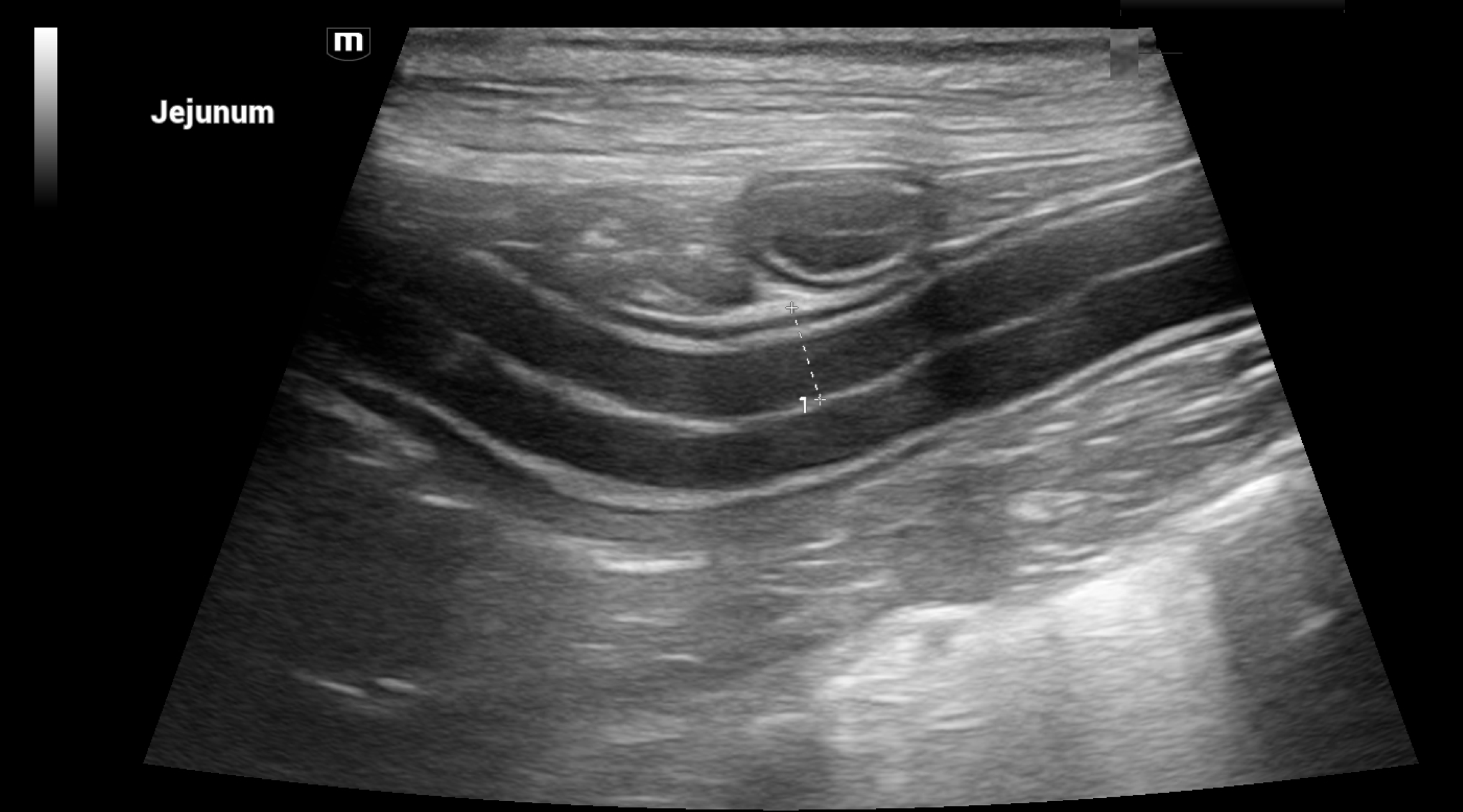
Ultrasound image of Jejunum
Ultrasound image of a section of jejunum with visible spiral-shaped intestinal folds.

正常與異常(增厚)腸道-診斷腸道疾病的關鍵。

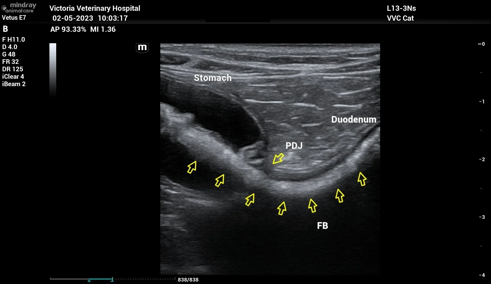
Ultrasound image of a patient's abdomen showing the stomach, duodenum, PDJ (pancreatic duct junction), and FB (foreign body), with areas marked by yellow arrows.

超聲波影像顯示貓吞食了線狀異物(繩子)。

心臟超聲波:用於檢查心臟。我們可以利用動態影像來辨識心臟內部的異常情況。

心臟瓣膜(二尖瓣)關閉不全的彩色血流影像(CFD)超聲波診斷

我們可以利用專用工具測量心臟內的血流與壓力,從而確定正確的治療方案,並監測治療效果:
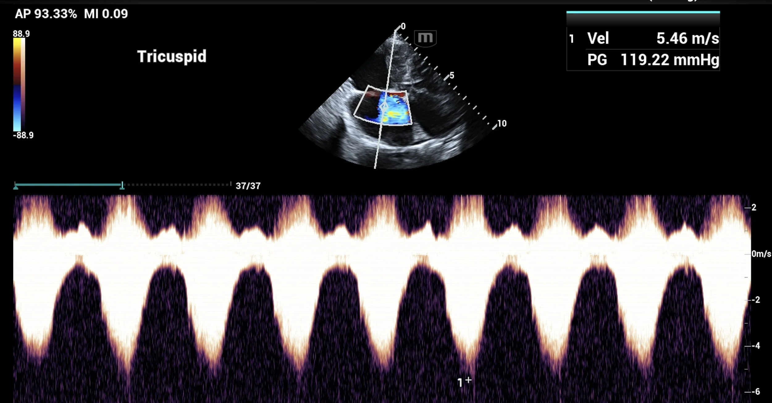
Ultrasound guide with Doppler color flow and spectral Doppler waveforms showing blood flow in the tricuspid valve.

犬肺動脈高壓的頻譜多普勒超聲波診斷

超聲波引導下診斷性採樣:我們可以利用超聲波引導對腫瘤等內部異常結構進行採樣,從而實現快速、準確的診斷。

超聲波引導下腹部腫塊採樣

懷孕診斷:有時我們會承擔一項非常令人愉快的任務,利用超聲波檢查是否有新的小狗或小貓孕育其中,並確認牠們的健康狀況!

一隻45天大的幼犬

內窺鏡

內窺鏡是一種透過光纖電纜連接到柔性鏡體上的攝影設備,用於觀察體內部情況。

在維多利,我們有多種適用於不同用途及解剖部位的內視鏡。

我們最大型的內視鏡用於胃腸內視鏡檢查,用於上消化道(食道、胃和小腸)和下消化道(結腸)內視鏡檢查。
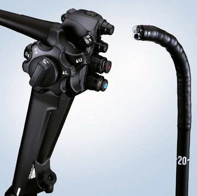
Close-up of a medical endoscope control handle with buttons and knobs, alongside a flexible black endoscope tube.

此內窺鏡可用於檢查這些部位是否有異常,並對異常組織進行活檢取樣。

An endoscopic view of the inside of a colon.

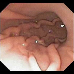
胃內窺鏡檢查

Microscopic image of tissue showing different cells and structures.

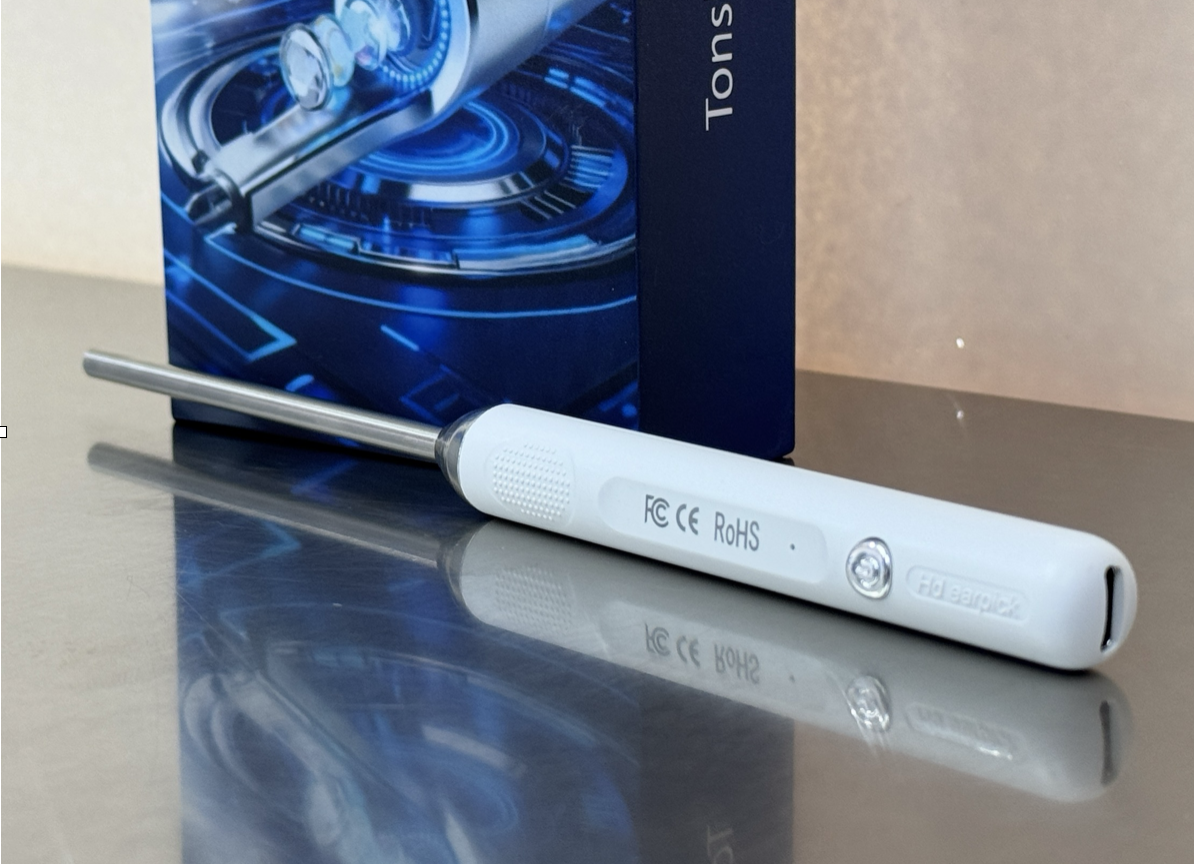
我們也提供一系列可與手機或平板電腦搭配使用的「便攜式」內窺鏡,適用於耳部和鼻部等較小部位的檢查;在診間使用時,還能讓客戶即時查看耳部檢查情況。
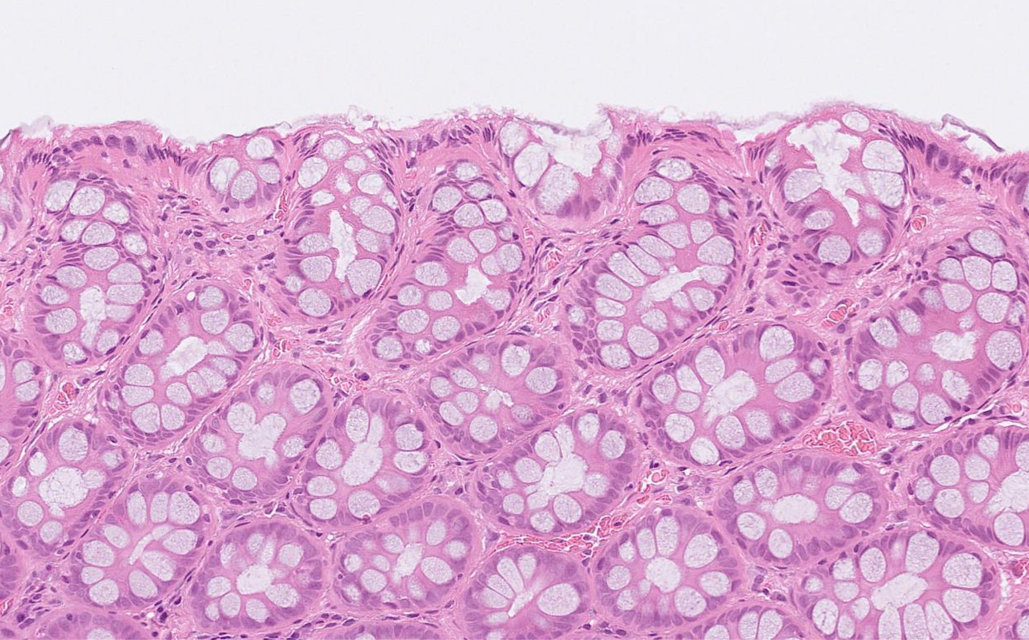
在顯微鏡下的結腸活檢樣本

此外,此內窺鏡還可用於定位並取出食道或胃內的異物。
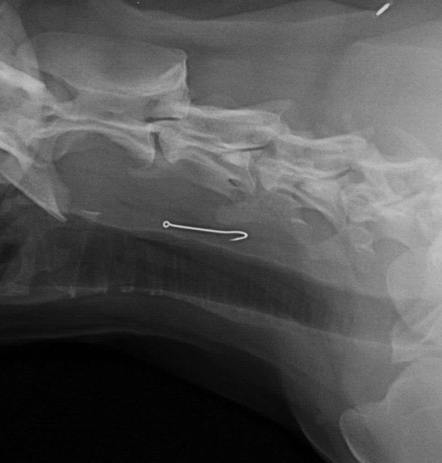
X-ray of a neck with a surgical hook visible.
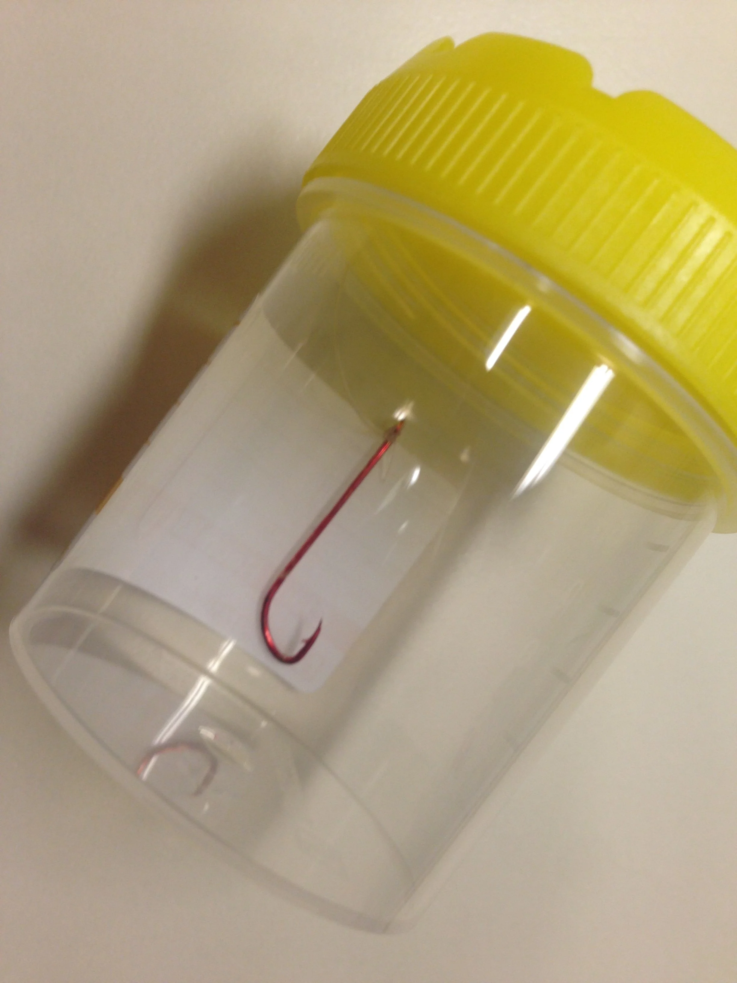
Close-up of a clear plastic container with yellow lid and a fishing hook inside.

被狗吞食的魚鉤,內窺鏡取出術前與術後的比較

我們還有尺寸更小、足以進入氣道的設備。這種檢查稱為支氣管鏡檢查;在病患接受麻醉的狀態下,它能讓我們觀察氣道內部,並從病變部位採集液體或組織樣本。
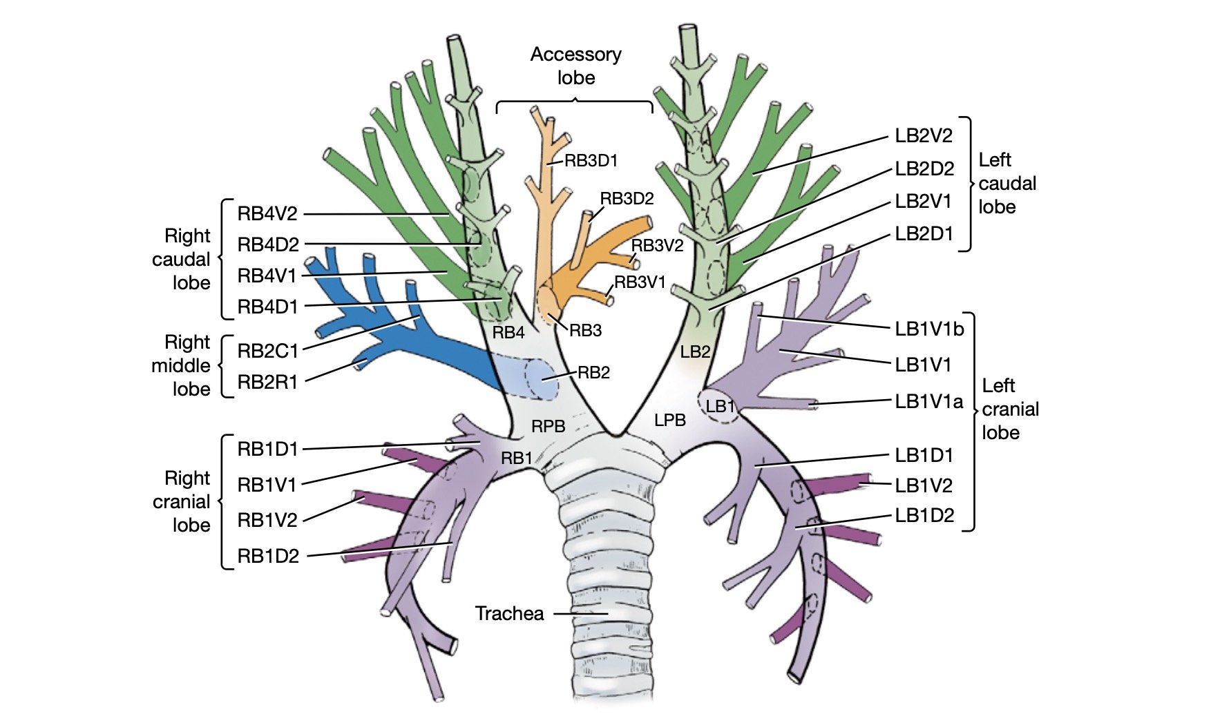
Diagram of the dog bronchial tree showing the right and left lungs, the trachea, and various branches labeled with their anatomical names, including lobes, bronchi, and segmental bronchi, color-coded for clarity.

犬隻氣道示意圖

麻醉狀態下主氣道的支氣管鏡檢查影片

A portable mini endoscope device lying on a reflective surface, with a blue box in the background.